Resorption is a pathological process in which hard tissues in the teeth, like dentin, cementum, and occasionally bone, are lost. Depending on where it is and the underlying causes, it has been classified into different categories;


Resorption is a pathological process in which hard tissues in the teeth, like dentin, cementum, and occasionally bone, are lost. Depending on where it is and the underlying causes, it has been classified into different categories;
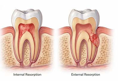
Internal root resorption is used to describe the resorption originating in the root canal within the tooth; while external root resorption is the term used to describe root resorption that originates on the external root surface. (Patel et al., 2023)
As per the ESE position statement, it has been classified further into; Internal resorption which could be inflammatory internal resorption or replacement internal resorption. External root resorption which could be inflammatory, replacement, surface, or cervical external resorption.(Patel et al.,2023)
- Internal Root resorption:
Where Resorption initiates from the pulp chamber or root canal on the inside of the tooth and extends from there. Loss of dentin and occasionally the adjacent firm tissues is its hallmark. Typically observed as a radiolucent area surrounding the pulpal cavity.
Its aetiology is not clear, as multiple causative factors has been suggested as; trauma, orthodontic forces and infection.
Pathogenesis:
Initiated by damage of the odontoblastic and the unmineralized predentine layer and exposing mineralized dentine to pulp tissue, causing odontoclasts to migrate to the site of injury and resorbing the changed root canal walls. (Andreasen & Andreasen, 1988).
Clinically:
Usually are asymptomatic, and could develop into irreversible pulpitis, advanced cases could show symptoms of pulpal necrosis. Some cases are associated in changes in color of the affected tooth.
Testing: Sensibility testing could be positive or negative depending on the pulpal status.
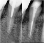
Radiographical signs: Internal resorption is viewed as a round-to-oval radiolucency (as in inflammatory types) in the root canal that is continuous with the canal wall, or in case of root fracture starting at the fracture line, or Mottled appearance (replacement internal resorption).
CBCT is recommended for cases of internal root resorption as a diagnostic aid. (Patel et al., 2014)
Management:
The main aim of the management is to clean the root canal system thus removing
The vital apical pulp tissue that sustains the resorption process. Thus clinical management includes full root canal treatment process with perforation repairs in case of occurrence as well surgical perforation repairs, Or extraction in cases tissue loss was unrepairable.
- External Resorption:
It is a pathological process in which the exterior surface of the root resorbs due to clastic cell activity, resulting in the loss of cementum, dentin, and, in some cases, bone. The most predisposing factors known are trauma and orthodontic forces. (Patel et al., 2022)
Pathogenesis:
According to (Fuss et al., 2003) it Includes an interaction of several biological processes:
Trauma and Mechanical Stress: Trauma can cause damage or hypoxia of the periodontal ligament, allowing osteoclasts and other clastic cells to reach and resorb the root surface.
Inflammatory Mediators: Periodontal infection or inflammation causes the release of cytokines and other pro-inflammatory mediators, which can trigger clastic cells to resorb tooth structure.
External resorption has been identified into(Patel et al., 2022) :
A/External Surface resorption:
Which is pressure-induced resorption and occurs on the external surface of the root, it is noninfective and self-limiting and stops with the removal of causative force.
Etiology: forces exerted due to impacted teeth, odontogenic cysts, orthodontic forces or trauma. (Levander & Malmgren, 1988).
Clinical Features: usually associated pulp is vital, and detected as an accidental radiographical finding.
Some delayed cases mobility, change in color could be associated
Radiographic feature:
As asymmetric loss of root surface neighbouring causative force (as cyst or impacted tooth), or as a blunt root surface incase of orthodontic causative forces.
Clinical Management: elimination of the causative factor, as for cases of orthodontic treatment a course either temporary pause of the treatment or termination of it in cases of significant resorption observed. (Mehta et al., 2017)
Follow-up and evaluation of stability: Active and stable (repaired) external surface resorption can be distinguished by the disappearance and re-establishment of the periodontal ligament space and lamina dura, respectively. (Sondeijker et al., 2020).
B/External Cervical resorption:
is initiated in the cervical region of the tooth, immediately beneath the epithelial attachment, and it is frequently aggressive.
Aetiology:
Multiple factors have been identified; such as trauma(as in cases of luxation forces), orthodontic treatment, internal bleaching procedures, surgical procedures such as periodontal surgeries, or certain systemic conditions.
Pathogenesis:
Develops through phases: as an invasion of the osteoclastic cells through the cementum, then the resorption phase where the lesion starts to resorb, consisting of fibrovascular tissues, then spreading circumferentially apico-coronally, in advanced cases a stage of reparative phase could be observed where bone-like tissue is deposited into the lesion.
It has been classified according to two systems, Patel and Heithersay.
Heithersay Classification of External Cervical Root Resorption: which relies on 2D radiographic imaging.
- Small lesion near the pulp with shallow dentine penetration
- Marked invasive lesion with minimal to no extension into the radicular dentin, penetrating near the coronal pulp chamber.
- Thorough invasion of dentin with tissue resorption reaching at least the coronal third of the root in addition to the coronal dentin.
- Extensive invasive resorption penetrating the coronal part of the root canal
Patel 3D classification (Patel et al., 2017): classified according to circumferential spread, hight, and extent of pulpal involvement, with the aid of 3D radiography and periapical radiography.
Hight: Circumefertial spread Pulp involvement
1-Hight to cementoenamel A <=90 d. confined to dentine
junction.
2-Cervical third B >90 to ≤180 P.probable pulp involve
- Middle third C >180 to ≤270
- Apical third D >270
Clinical Signs and Symptoms:
Usually reflect according to the above classifications, yet early lesions are asymptomatic, and usually detected as accidental clinical/radiographical findings. More progressive classes are usually presented as periodontal lesions or pulpitis, or in-case of granulation tissue reflected as pink discoloration through the tooth cervically. clinical probing often reflects a scratchy tactile sensation or results in bleeding.
Radiographically:
Depending on the pathological phase reflects as either radiolucency, mottled appearance, or ragged. As they don’t have a classic characteristic appearance.
Cone beam computed tomography aids in the diagnosis and classification of the lesion thus provides more clear guidance in the clinical management of the lesion.\
Treatment:
The clinical management depends on the extent of the resorptive lesion, and mainly aiming at removal of the resorptive tissue and restoration of the defect.
Including external access with a surgical approach including raising a flap to allow removal of the resorptive tissue, with or without root canal treatment depending on the stage of the lesion. An internal clinical approach where mechanical removal of the tissue is done (non-surgically) with or without root canal treatment depending on the extent of the lesion to the pulp or ease of restoration application in the lesion clinically.
Resection of the involved root or Extraction; In cases where the lesion is too advanced, and the prognosis of the tooth is poor.
Materials of choice to restore the lesion were calcium silicate-based types of cement, composite resins, or glass ionomer restorations, depending on the extent of the lesion clinically. (Bardini et al., 2023)
Prognosis and follow-up:
The outcome of the intervention and the size of the resorptive lesion determine the prognosis. While late lesions with substantial structural damage may need to be extracted, early-stage lesions typically have a good prognosis.
Prosthodontic replacement always has to be discussed with patients in advanced ESR cases and posterior teeth.
C/ External inflammatory resorption-infection related:
Occurs in cases where Infected necrotic pulp damages the root canal microbiota and the protective pre-cementum layer on the root surface, could be seen related to severe luxating injuries as avulsion.
Clinically:
The majority are asymptomatic and detected as accidental findings, yet symptoms of irreversible pulpitis, with a negative response to pulp sensibility testing.
Radiographical feature:
Loss of lamina dura, ragged root end, and shorter root are key features in 2D radiography, 3D imaging is advised to detect adequate extent and perforations.
Treatment:
Aimed to remove the causative factor, by complete root canal treatment with proper disinfection.
Extraction is the option for cases where severe destruction occurred.
Prognosis:
Following ideal root canal treatment, healing and arrest of the resorption process occurs.
D/ Replacement Resorption (Ankylosis):
In cases of severe resorption, a replacement resorption results from progressive replacement of the tooth structure with bone.
Aetiology:
Usually occurs following severe trauma incidents, especially avulsion and reimplantation of teeth and where extensive damage to the periodontal ligament is related to the tooth.
Clinical signs and symptoms:
Percussion produces a key metallic sound, and often becomes non-responsive to sensibility tests as result of subsequent nerve damage.
Possibly to appear infra occluded, as in cases where the history of trauma has been associated in developing dentintion.
Radiographic appearance:
2D periapical radiographs show a less distinct outline of periodontal ligaments which could disappear over time completely. CBCT is advised to asses the extent or degree of the replacement resorption associated.
Treatment:
The choice of treatment depends on many factors, such as the age of the patient’s development, the extent of the resorption associated, and the location of the affected tooth.
- Extraction and implant-as in cases where its aesthetically non-pleasing appearance, as well in cases where orthodontic management is planned as ankylosed teeth will not respond to orthodontic movement.
- De-coronation, where removal of the crown only allows bone preservation for either future implant planning or future possible root resorption and replacement to bone.
- Monitoring. Annual periodic reviews.
- Composite buildup for infra-occluded teeth to allow a pleasing aesthetic outcome.
Prognosis:
In cases of young patients, with developing dentition or late detection ankylosed teeth with replacement resorption are generally of poor prognosis.
D/Transient apical breakdown
Defined as temporary radiolucency or disintegration at the apex of a tooth, as a result of trauma, most frequently observed in young, developing teeth.
Aetiology:
Following trauma concussion or subluxation traumas; temporary disruption of apical blood supply occurs followed by a repair process, which is reflected as this form of breakdown.
Clinical signs and symptoms:
Usually symptomatic, sometimes delayed sensibility response is associated.
Radiographical appearance: Widening of periodontal ligament space.
Management:
Depends on the risk of pulpal involvement following monitoring, as usually it’s a self-limiting case.
Conclusion:
Identification of the causative factor is the key point for proper management, with the aid of CBCT imaging and proper history taking higher prognosis has been associated with root resorption cases.
References:
Patel, S., Krastl, G., Weiger, R., Lambrechts, P., Tjäderhane, L., Gambarini, G., & Teng, P.-H. (2023). ESE position statement on root resorption. International Endodontic Journal. https://doi.org/10.1111/iej.13916
Fuss, Z., Tsesis, I., & Lin, S. (2003). Root resorption – diagnosis, classification and treatment choices based on stimulation factors. Dental Traumatology, 19(4), 175–182. https://doi.org/10.1034/j.1600-9657.2003.00192.x
Andreasen, F. M., & Andreasen, J. O. (1988). Resorption and mineralization processes following root fracture of permanent incisors. Dental Traumatology, 4(5), 202–214. https://doi.org/10.1111/j.1600-9657.1988.tb00323.x
Patel, S., Durack, C., Abella, F., Roig, M., Shemesh, H., Lambrechts, P., & Lemberg, K. (2014). European Society of Endodontology position statement: The use of CBCT in Endodontics. International Endodontic Journal, 47(6), 502–504. https://doi.org/10.1111/iej.12267
Patel, S., Saberi, N., Pimental, T., & Teng, P. (2022). Present status and future directions: Root resorption. International Endodontic Journal. https://doi.org/10.1111/iej.13715
Levander, E., & Malmgren, O. (1988). Evaluation of the risk of root resorption during orthodontic treatment: A study of upper incisors. The European Journal of Orthodontics, 10(1), 30–38. https://doi.org/10.1093/ejo/10.1.30
Mehta, S. A., Deshmukh, S. V., Sable, R. B., & Patil, A. S. (2017). Comparison of 4 and 6 weeks of rest period for repair of root resorption. Progress in Orthodontics, 18(1). https://doi.org/10.1186/s40510-017-0173-1
Sondeijker, C. F. W., Lamberts, A. A., Beckmann, S. H., Kuitert, R. B., van Westing, K., Persoon, S., & Kuijpers-Jagtman, A. M. (2019). Development of a clinical practice guideline for orthodontically induced external apical root resorption. European Journal of Orthodontics, 42(2), 115–124. https://doi.org/10.1093/ejo/cjz034
Patel, S., Foschi, F., Mannocci, F., & Patel, K. (2017). External cervical resorption: a three-dimensional classification. International Endodontic Journal, 51(2), 206–214. https://doi.org/10.1111/iej.12824
Bardini, G., Cristiano Orrù, Ideo, F., Venkateshbabu Nagendrababu, Dummer, P., & Cotti, E. (2023). Clinical management of external cervical resorption: A systematic review. Australian Endodontic Journal, 49(3), 769–787. https://doi.org/10.1111/aej.12794
Book Now
It’s easy and free!